
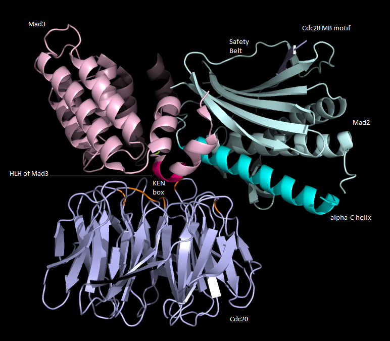
Overall Structure of the MCC:
· Mad2, Mad3 and Cdc20 were co-expressed inside an insect cell to create the yeast MCC when they formed a complex.
· Contained Cdc20 with complete functional domains, Mad2 present as C-Mad2 and Mad3 was added but it was cleaved after its tetratricopeptide repeat (TRP) domain, which is a structural motif composed of antiparallel α-helices, so that it lacked its carboxy-terminus D box [2].
 |
| Figure 1. Overall structure of the MCC animated gif. |
The crystal structure was solved to a resolution of 2.3 Å:
· Side chains of the amino acid residues should be clearly visible. May be depressions in the rings of aromatic amino acids.
· The structure at this resolution shows that Cdc20 (blue), Mad2 (light cyan) and Mad3 (light pink) form a triangular heterotrimer as shown in Figure 1.
· Mad3 coordinates the overall organisation - forms many interactions between Mad2 and Cdc20.
· Mad2 and Cdc20 interact mainly through the sequestering of the Mad2-binding (MB) motif on Cdc20 by safety belt of Mad2.
· Mad3 is a core of contiguous TRP superhelix - 3 TPR motifs flanked by capping α-helices. Mad3 has a highly conserved amino-terminal sequence which contains its amino-terminus KEN box essential for MCC assembly in a helix-loop-helix (HLH) motif.
· HLH motif is essential for the MCC formation - simultaneously binds Mad2 and Cdc20, and in doing so it orientates the KEN box towards its receptor on the WD40 domain of Cdc20.
· Mad3 also contacts Mad2 and Cdc20 with its TPR motifs [2].
 |
| Figure 2: Overall structure of the MCC with labels. |
Figure 2:
- shows the overall structure of the MCC, but with each of the residues highlighted according to their B factor.
- B factor is a measure of the degree of flexibility of a given residue. The residues that are least flexible are highlighted dark blue, and any red residues indicate a highly flexible residue.
- This structure of the MCC shows mostly blue residues in the Mad3 and Cdc20 regions, showing that there is not much flexibility in this region and so this structure is likely to be correct. Mad2 has some residues that are coloured orange, indicating that they are relatively flexible. Mad2 undergoes transitions from its open to closed states, and perhaps this is why these regions are particularly flexible.
 |
| Figure 3. the MCC coloured according to B factors. |
Mad2:
· Structure reveals C-Mad2 conformation allows it to interact with the MB motifs on Cdc20 and Mad1. C-Mad2 is the only conformation of Mad2 that enables it to recognise Mad3.
· Mad2 interacts with the HLH motif of Mad3 through its α-C helix and β8’-β8’’ hairpin loop.
· These interactions of Mad2 resemble those interactions between C-Mad2 and MAD2L1-binding protein, and the interactions of C-Mad2 with O-Mad2 in an asymmetric dimer.
· The β8’-β8’’ region of Mad2 undergoes a great conformational change when Mad transitions from open to closed states, and the participation of this hairpin in the interaction to the HLH motif of Mad3 indicates that Mad3 binds to the C-Mad2 exclusively [2].
Good use of pictures and animations, the animation allows the reader to get a feel for the 3D shape of the complex and the picture, with its labels, clearly demonstrates the important structural details.
ReplyDelete